Home | Industries | Laboratory| PVDF Western Blotting Membrane

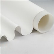
PVDF Western Blotting Membrane

Immunoblotting test is also known as western blot, which is a hybrid technology combining high-resolution gel electrophoresis and immunochemical analysis.

Western blot has the advantages of large analytical capacity, high sensitivity and strong specificity. It is the most common method to detect the characteristics, expression and distribution of proteins, and is widely applicated in:


• Study on Gene Expression at Protein Level
• Antibody Activity Test
• Early Diagnosis of Disease


Advantages

• Smooth and flat surface, not easy to curl
• High mechanical strength, easy to be stripped and reprobed
• Evenly distributed pore size, high protein mobility, clear results
• High sensitivity to ensure successful detection of low abundance protein


Application

• Western Blot

• Adsorption Analysis

• Amino Acid Analysis

• N-terminal Protein Sequencing

• Spot and Slit Printing Detection

• Glycoprotein Coloration

• Lipopolysaccharide Analysis


Performance Comparison of Immunoblotting Membrane

Membrane MaterialNCPVDF
Protein binding capacity μg/cm2LowHigh
Mechanical strengthFragileDurable
Chemical compatibilityLimited

Compatible with most organic solvents,
acids and weak bases

Whether activation is requiredNoMethanol activation required
Detection

Chemiluminescenc
Fluorescence detection
Conventional dyeing

Chemiluminescence
Fluorescence detection
Conventional dyeing
Coomassie blue staining

ReprocessingUnsuitableSuitable
Heat resistancePoorGood


Comparison of PVDF Transfer Membrane


Company / ModelM CompanyT Company

Cobetter
TS2590L

Cobetter
TS2590H

Application

Common
immunoblotting 

Common
immunoblotting 

Especially suitable for low
abundance protein detection

Common
immunoblotting 

MaterialPVDFPVDFPVDFPVDF
Pore Size / μm0.450.450.450.45
Thickness / μm12712791115
Test Method

Chemiluminescence
 Substrate Coloration

Results of 45kDa
Proteins

PVDF western cbt 00 (1).pngPVDF western cbt 00 (12).pngPVDF western cbt 00 (11).pngPVDF western cbt 00 (10).png

Results of 35kDa
Proteins

PVDF western cbt 00 (9).pngPVDF western cbt 00 (8).pngPVDF western cbt 00 (7).pngPVDF western cbt 00 (6).png

Results of Proteins
less than 30kDa

PVDF western cbt 00 (5).pngPVDF western cbt 00 (4).pngPVDF western cbt 00 (3).pngPVDF western cbt 00 (2).png


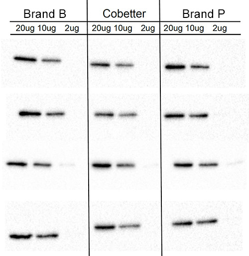
According to the chemiluminescence test results, the 0.45μm hydrophobic PVDF transfer membrane developed by Cobetter can achieve similar protein transfer effect as competitor’s products.

The 0.2 micron model is under development.


Performance Comparison of Immunoblotting Membrane

Membrane MaterialNCPVDF
Protein binding capacity μg/cm2LowHigh
Mechanical strengthFragileDurable
Chemical compatibilityLimited

Compatible with most organic solvents,
acids and weak bases

Whether activation is requiredNoMethanol activation required
Detection

Chemiluminescenc
Fluorescence detection
Conventional dyeing

Chemiluminescence
Fluorescence detection
Conventional dyeing
Coomassie Blue Staining

ReprocessingUnsuitableSuitable
Heat resistancePoorGood


Strong protein adsorption capacity and signal low background make hydrophobic PVDF membrane perform better than other materials. Especially under rigorous conditions, such as when organic, acid and alkali solvent occurs. PVDF has better sensitivity and less brittle than nitrocellulose as well. 


In addition, PVDF membrane is reusable without a loss of sensitivity or increased background.  


Comparison of performance between Cobetter membrane and other brands. 


Figure1. 


Specification

  Pore size  

   0.2μm

 Standard width 

   270mm  (can be changed based on requirement )

  Length

   Based on the requirement from clients


Comparison  between Cobetter PVDF western blotting membrane with other brand membranes

PVDF-Western-Blotting-Membrane-1.jpg


Ordering Information

ModelPore SizeLength (mm)Width (mm)
TS2590L0.45 μm2603300

High sensitivity model, suitable for detection of low abundance protein..

TS2590L297210
TS2590L210148
TS2590L200200
TS2590L150150
TS2590L8470
TS2590H0.45 μm2603300General model, relatively high sensitivity, suitable for detection of high abundance protein
TS2590H297310
TS2590H210148
TS2590H200200
TS2590H150150
TS2590H8470


* More sizes can be customized.

DOWNLOAD

Contact Form